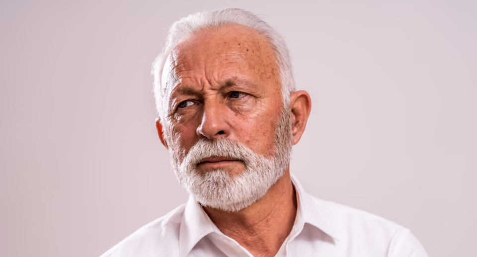
Ptosis, or upper eyelid drooping, is a common condition that can significantly impact the superior visual field, making everyday tasks like reading, driving, or navigating obstacles more difficult. When ptosis becomes visually significant, eye doctors rely on ptosis visual field testing, particularly the Superior 36 visual field test, to document functional impairment and determine whether eyelid surgery is medically necessary.
This guide is built for understanding ptosis. You’ll learn how ptosis obstructs peripheral vision, why eyelids are taped during visual field exams, and how modern tools like the Carrot headset streamline taped and untaped visual field testing for insurance reimbursement and ptosis surgery qualification.
What Is Ptosis?
Ptosis (blepharoptosis) refers to the drooping of one or both upper eyelids. Even mild drooping can block the superior field of vision, creating noticeable functional limitations. Some patients experience difficulty seeing road signs, reading, or maintaining eye contact, all due to eyelid obstruction.
Ptosis is often misunderstood as purely cosmetic, but in many cases it is a functional vision problem. A ptosis eye test or ptosis visual field test provides objective confirmation of whether the eyelid position is reducing measurable vision.
Types of Ptosis
Ptosis may be congenital or acquired. Common forms include:
- Aponeurotic ptosis – due to tendon stretching (most common)
- Myogenic ptosis – associated with muscle weakness or myasthenia gravis
- Neurogenic ptosis – caused by nerve dysfunction
- Mechanical ptosis – excess weight on the lid from lesions or swelling
- Traumatic ptosis – injury affecting lid muscles or nerves
- Pseudoptosis – overhanging skin simulates lid droop
All forms can cause superior visual field loss, often evaluated through ptosis visual field testing.
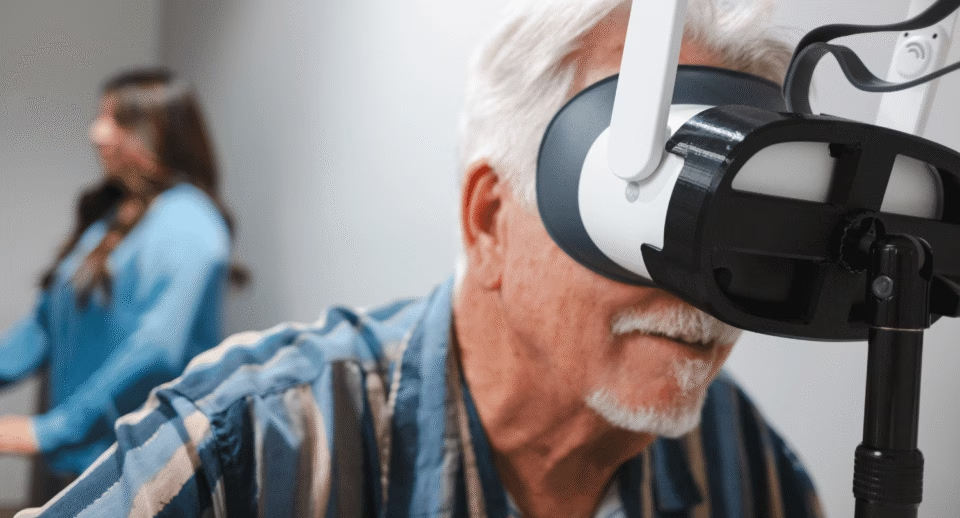
Why Does the Eye Doctor Tape Eyelids for Peripheral Vision?
Patients frequently ask: “Why does the eye doctor tape eyelids for peripheral vision tests?”
During a taped and untaped visual field test, clinicians compare:
- Untaped visual field results – shows true functional loss from ptosis
- Taped results – simulate the visual field after surgery
This comparison provides the evidence needed for:
- Determining medical necessity
- Demonstrating functional improvement
- Meeting insurance and Medicare requirements for eyelid surgery
Accurate documentation is essential, especially for those researching how to qualify for eyelid surgery or navigating insurance approval.

The Superior 36: A Targeted Peripheral Vision Test for Ptosis
The Superior 36 visual field test, also known as the Superior 36 point suprathreshold test, is the preferred visual field test for ptosis, blepharoplasty qualification, and documenting superior field loss.
How the Superior 36 Visual Field Test Works
Understanding the Superior 36 visual field test supports understanding ptosis. This test:
- Uses 6 dB suprathreshold stimuli
- Tests points up to 52 degrees in the superior visual field
- Each point is tested once and repeated if missed
- Performed taped and untaped to determine true eyelid obstruction
- Faster and more targeted than full threshold exams like the Humphrey 24-2
This streamlined approach makes it the ideal visual field test for eyelid surgery and visual field test for blepharoplasty.
Eye doctors using Humphrey may compare this to the Humphrey visual field test for blepharoplasty, but the Superior 36 was designed specifically for functional assessment of ptosis.
Challenges in Ptosis Visual Field Testing
Accurate ptosis visual field test interpretation can be affected by:
- Poor fixation
- Delayed patient responses
- Variability in eyelid taping method
- Dry eye, media opacity, or pupil size differences
- Patient fatigue
Because insurance decisions rely on these results, clinicians must ensure standardized, repeatable testing, something VR-based devices can significantly improve.
From Screening to Surgery: Making Treatment Decisions
When visual field tests show eyelid overhang interfering with vision, surgeons evaluate whether ptosis repair is appropriate. Insurance companies typically require:
- Clear superior visual field loss on untaped testing
- Significant improvement in taped results
- Patient-reported functional impairment
- Documentation with photos, plus the ptosis field vision test
Some patients search online for “how to fail a visual field test for eyelid surgery” or “how to fail a field vision test for ptosis,” but clinicians know the goal is well-documented accuracy, not manipulation. Proper taping methods and standardized testing ensure reliable, medically appropriate results.
In mild cases, nonsurgical options like oxymetazoline (Upneeq) may be considered.
Understanding Ptosis Visual Field Test Reports (Superior 36)

Interpretation focuses on comparing taped vs. untaped findings.
Typical findings in a ptosis visual field defect:
- Missed points clustered in the upper field
- Reduced superior sensitivity due to eyelid obstruction
- Improved detection of stimuli when the eyelid is taped
Indicators supporting surgery:
- ≥12 degrees or ≥30% improvement in the superior field when taped
- Documented limitations affecting daily activities
- Photo evidence of ptosis obstructing the visual axis
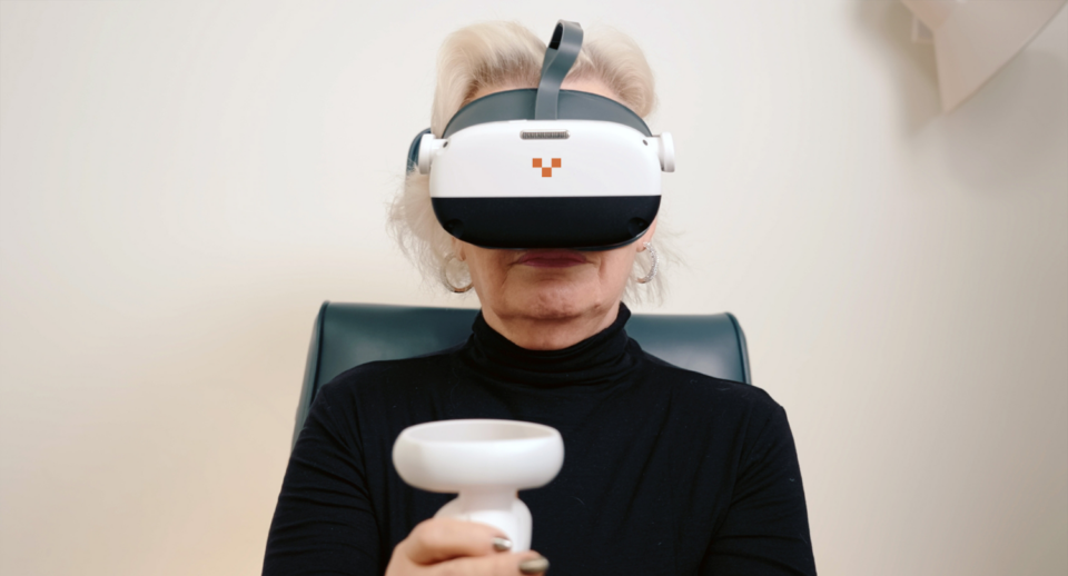
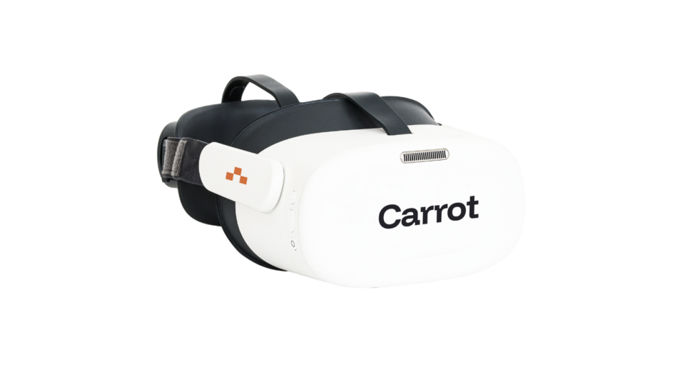
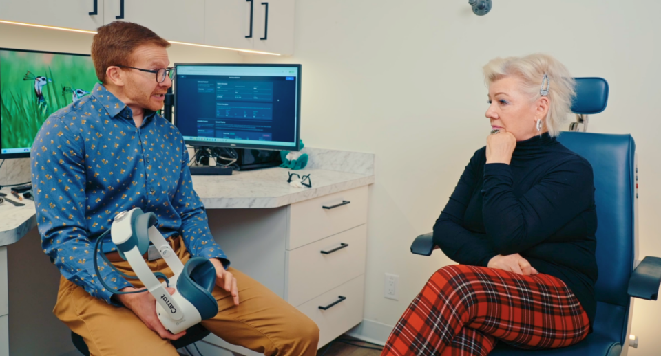
Carrot — The Modern Way to Perform Superior 36 Testing
The Carrot headset simplifies and standardizes the Superior 36 visual field test, helping practices meet insurance and Medicare requirements with ease.
Why Carrot is the Best Tool for Ptosis Visual Field Testing
- Fully supports taped and untaped visual field protocols
- Validated Superior 36 visual field test built into the workflow
- Portable VR design improves patient comfort and accuracy
- Faster than traditional bowl perimeters
- Produces clean results for insurance documentation
- Ideal for oculoplastics, general ophthalmology, and high-volume clinics