While the need for eye care services is on the rise, the eye care industry grapples with a shortage of ophthalmologists and ophthalmic technicians. Consequently, medical optometrists have emerged as key players in bridging this gap and delivering comprehensive care. Various technological innovations, such as virtual visual field testing, are instrumental in helping optometrists fulfill this crucial role.
In this article, we will discuss how medical optometrists can maximize visual field utilization to improve patient care and provide a more efficient experience.
Driving and Visual Field Testing
Utilizing visual field testing becomes a crucial tool in enhancing driver safety, especially considering that nearly nine out of ten adult Americans engage in driving, with over 20% of them aged 65 or older, an age group expected to expand. This growing demographic is at increased risk of ocular diseases that can compromise vision. Given that car accidents stand as a leading cause of death, and older drivers contribute to 13% of fatal traffic crashes, placing a priority on eye health is paramount.
To tackle this issue, regular eye examinations, including visual field testing such as the Esterman exam, become essential for elderly drivers. The Esterman exam offers a comprehensive assessment, surpassing the peripheral vision prerequisites for (most) state licensing exams, typically set at 120 degrees. This advanced technology provides a more in-depth understanding of visual capabilities.
When visual field testing reveals that a patient falls below state driving requirements, it sparks a challenging conversation. Safeguarding the independence of elderly individuals, closely linked to their ability to drive, takes precedence. Optometrists frequently use the Esterman visual field exam to educate patients, presenting and explaining the results to help them understand the limitations of their vision and make informed decisions about driving safety.
Visual Field Testing in Oculoplastics
Tests like the Superior 36 are valuable in directing oculoplastics referrals, covering a range from cosmetic procedures to eye diseases. Oculoplastic surgeons value the visual field results obtained prior to referrals.
In cases involving blepharoptosis or dermatochalasis, understanding the extent of impairment in the superior field of vision is crucial. Visual field testing becomes indispensable in assessing the degree of impairment. Testing can be conducted in the patient’s natural state and repeated during lid taping or holding to simulate the conditions of a surgical procedure.
In the evaluation of medical eye conditions such as thyroid eye disease, visual field testing is helpful for assessing lid position and identifying potential issues. Additionally, it provides insights into the status of the optic nerve, a critical aspect as proptosis progresses.
Glaucoma and Visual Field Testing
Visual field testing is a crucial tool for tracking glaucoma progression and evaluating treatment efficacy, serving both glaucoma suspects and diagnosed patients.
Visual field testing provides insights into our patients’ performance that extend beyond structural changes revealed by OCT. While OCT is pivotal for assessing structural alterations, understanding patients’ visual status remains integral.
Opting for a 24-2 format widens the field under examination, offering a comprehensive view. On the other hand, a 10-2 format delves into the central field, where early manifestations of disease, particularly retinal ganglion cell loss, become apparent.
For optometrists, two distinct approaches prove beneficial
- Glaucoma Suspects: Conduct an annual visual field 24-2 test on the patient and rely on OCT technology to determine the need for more frequent testing.
- Glaucoma-Diagnosed Patients: In newly treated patients, perform three to five field tests to establish a predictor of progression analysis. Data collection can be more aggressive, with tests conducted at three- or six-month intervals. Depending on the disease’s severity, alternate between the 24-2 and the 10-2 if specific points in the center area fixation of a 24-2 show abnormalities. The 10-2 aids in discerning the extent of the defect.

Visual Field Testing for Patients Taking Plaquenil
Plaquenil is a medication commonly used to treat autoimmune diseases and inflammatory conditions such as Sjogren’s disease, rheumatoid arthritis, and lupus. While it didn’t cause much concern in the past, patients are now using it for 10-20 years. The toxicity effect of the medication can be a concern, causing rheumatologists and primary care providers (PCPs) to refer their patients to optometrists.
Before starting Plaquenil, a patient can get visual field testing, color vision testing, and a baseline macular scan. Continual testing is conducted annually thereafter until they reach a certain number, typically between five and seven years of using the medication. The patient’s weight and dosage can play a role in how soon concerns about toxicity occur.
Once the patient reaches the designated timeframe, optometrists are recommended to conduct tests at six-month intervals. Vision loss resulting from Plaquenil toxicity may be irreversible, and progression can persist even after discontinuing the medication. Hence, proactive identification becomes crucial in managing potential risks.
Visual Field Testing for Patients with Diabetes and Macular Degeneration
Macular edema associated with diabetes, wet age-related macular degeneration (AMD), and geographic atrophy (GA) affects the foveal tissue in the macula, potentially disrupting your patient’s central vision.
Depending solely on visual acuity testing may not offer a comprehensive understanding of these conditions. Therefore, incorporating a visual field test becomes instrumental in pinpointing specific areas of visual gaps for a more thorough assessment.
Visual Field Testing for Migraine Patients
Migraines can manifest with diverse visual symptoms, including temporary vision loss, auras, flashes of light, and hemianopsia. As migraines are vascular events, the visual system typically recovers as vascular stress alleviates and blood flow returns to normal. However, for individuals experiencing these visual events for the first time, there can be a tendency to confuse them with stroke symptoms.
In diagnosing migraines and eliminating other potential causes of vision loss, visual field testing is invaluable, especially with 30-2 and pupilometry. When exploring neurological aspects, the visual field is a crucial tool for understanding how the visual pathway processes vision. For the broadest scan, the 30-2 test is the preferred choice.
Experts also advocate for incorporating a color vision test to assess nerve and macular function, along with a pupilometry exam to document pupil size.
Headaches can signal underlying concerns, ranging from tumors to aneurysms. Establishing robust baseline data for such patients is imperative for effective monitoring and anticipating future events.
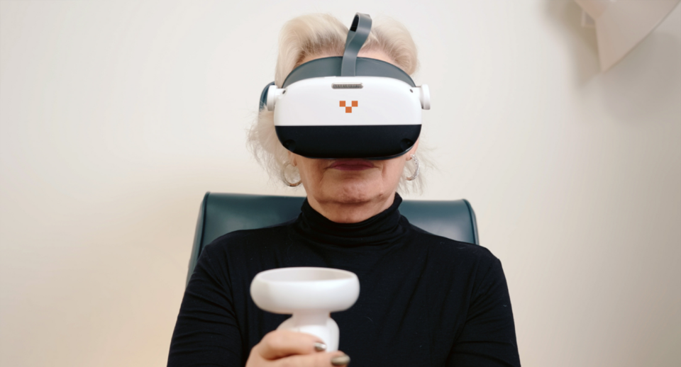
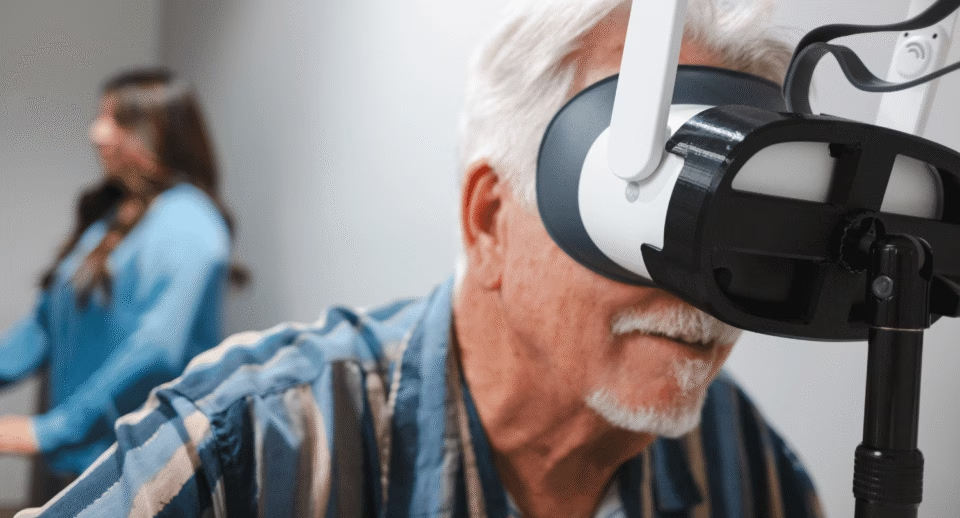
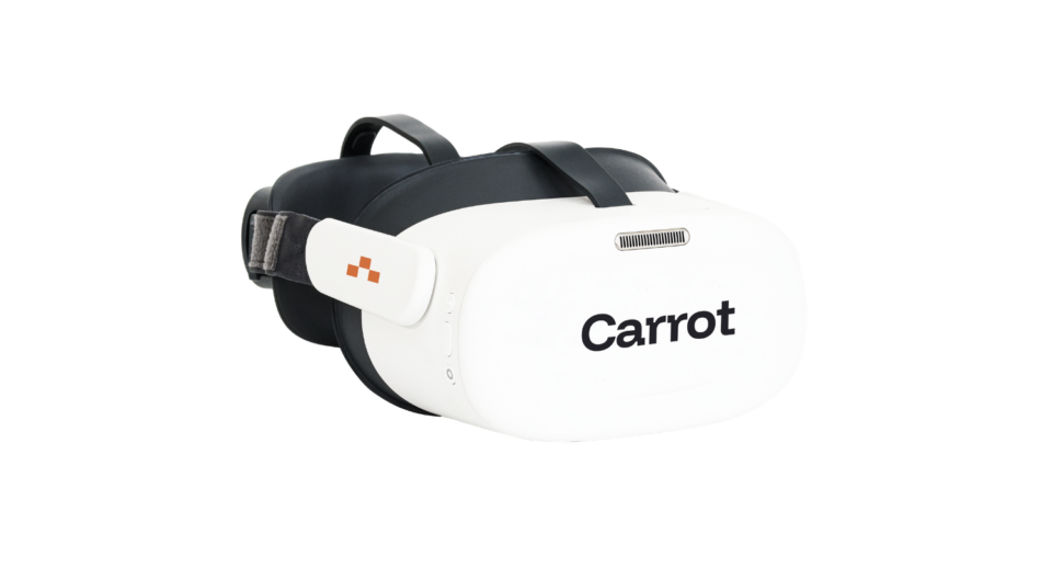
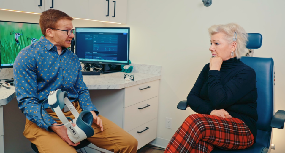
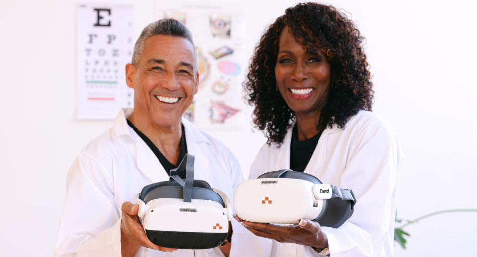
Using Carrot for Efficient Testing
Carrot provides a range of exams billable under four CPT codes, covering visual field exams for thresholding (24-2, 10-2, 30-2), screening (N-30 and C-40), ptosis (superior 36), and driving (Full Field 120, Esterman, Binocular Esterman). Additionally, it encompasses tests for color vision, foveal threshold, pupilometry, and kinetic (Goldmann).
The visual field testing offered by Carrot emerges as a valuable tool for evaluating various eye conditions. Medical optometrists find it particularly instrumental in managing conditions such as glaucoma, diabetes, macular degeneration, and migraines. This testing aids in both detecting and monitoring the progression of vision loss, serving as a means to educate patients about their specific eye conditions.